Ankle Case 10 Diagnosis
Weber B Fracture
Diagnosis
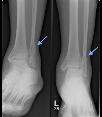
Standard three-view plain radiograph ankle series should be obtained which includes AP, lateral, and mortise (an AP view shot in 10-20° internal rotation) views. The fibular fracture line should be identified in relation to the ankle mortise. Fibular fractures at the mortise are Weber B. Weber B are often spiral fractures and frequently extend superiorly and laterally. If the spiral line originates at the mortise, as it commonly does, and spirals proximally, it is a Weber B, not a Weber C, fracture.
The joint space should be uniform all around the talus on the mortise view. The “medial clear space” between the medial malleolus and talus should be ≤ 4 mm. Radiographic signs of ankle instability/syndesmotic injury (details of which are beyond the scope of this case) include abnormalities of tibiofibular overlap and tibiofibular clear space.
Stability can be assessed without obtaining CT or MRI. In Weber B fractures, stability depends on whether the tibiofibular syndesmosis is intact or only partially torn, if the deltoid ligament is torn, and if there is medial malleolus fracture. Weber B fractures with medial malleolar fractures can be determined to be unstable from the initial x ray. When there is unstable deltoid ligament injury, there is typically widening of the MCS on the mortise view. However, some deltoid injuries may not show this widening on the standard mortise view. For this reason, for those with medial tenderness on exam and no evidence of MCS widening on standard x-rays, a stress view x-ray helps assess stability. To obtain this view, the physician stabilizes the affected lower leg with one hand proximal to the injury while the other hand grasps the great toe to evert the foot while an AP view is obtained. If the medial clear space is >4 mm on the stress view, a deltoid ligament injury can be assumed and the ankle is deemed unstable.