Ankle Case 11 Background
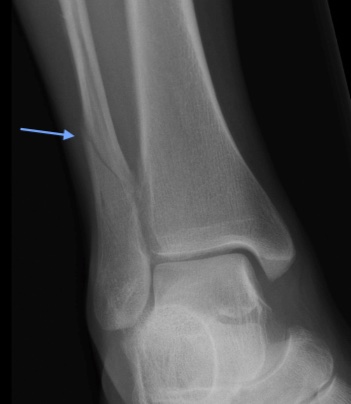
Weber C Fracture
Background
The ankle joint is made up of the articulation of the distal tibia, distal fibula, and the talus bone. Within this joint, the ankle mortise (think “recess”) is comprised of the articular surfaces of the distal tibia and fibula and houses the body of the talus within it. Additionally, there is a distal tibiofibular syndesmosis– the fibrous joint where the tibia and fibula are held together by several ligaments– as well as medial (i.e. deltoid)and lateral ligaments. Together, these structures form a ring and are responsible for providing stability to the ankle joint.
Ankle fractures make up 9% of all fractures. Distal fibula fractures (lateral malleolar fractures) are the most common. While there is no gender predominance for ankle fractures when compared over a lifetime, men have higher rates of distal fibular fractures at younger ages while women more often experience these fractures at older ages. In younger patients, high-energy trauma (i.e. MVCs, sports injuries) is more often the cause of fracture whereas in older patients, low-energy trauma (i.e. simple fall down a step) can cause distal fibular fractures.
Several classification systems exist to help assess ankle stability and guide treatment of distal fibular fractures. The most common system, the Weber classification (or Danis-Weber classification) system, is based on radiographic views of the ankle and differentiates lateral malleolus fractures based on the relation of the distal fibula fracture line to the ankle mortise:
- Weber A: fracture line occurs below, or distal to, the ankle mortise
- Weber B: fracture line originates at the level of the ankle mortise (can spiral superiorly/laterally) - the most common type
- Weber C: entire fracture line occurs above, or proximal to, the ankle mortise - the least common type.