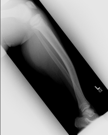
Bone Lesions Case 5 Diagnosis
Rickets
Diagnosis
 Clinical suspicion prompts an evaluation with X rays and laboratory tests.
These patients will have low calcium, phosphate, and vitamin D levels, with elevated alkaline phosphatase and PTH. Check renal function and all forms of Vitamin D..
X rays of the wrist, femur, and leg should be evaluated. The wrist will show changes first since it has the most rapid growth rate. Widening of the growth plate is a typical early change. This progresses to cupping, cortical spur formation, and thinning of the cortices of long bone. Later in the course of the disease, especially in osteomalacia, pseudo-fractures in the femoral neck can be seen. These are lucencies that mimic fractures, but are bilateral and not true fractures.